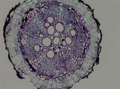
Fluorescence microscope a wonderful journey to explore the microscopic world
Basic principles of fluorescence microscopes
The working principle of fluorescence microscopes is based on the phenomenon of fluorescence. When certain substances (such as fluorescent dyes, quantum dots or fluorescent proteins) absorb light of a specific wavelength (excitation light), they will be in a high-energy state for a short time, and then release energy in the form of longer wavelength light (fluorescence) to return to a low-energy state. Fluorescence microscopes selectively excite fluorescent substances in samples by precisely controlling the wavelength of excitation light, and use optical systems to collect and amplify these fluorescence signals, ultimately presenting high-contrast images.
Types and development of fluorescence microscopes
With the continuous advancement of technology, fluorescence microscopes have developed into a variety of types to meet different research needs. Among them, the most common ones include epifluorescence microscope, confocal fluorescence microscope, multiphoton fluorescence microscope and super-resolution fluorescence microscope.
Confocal fluorescence microscope: Through laser scanning and pinhole filtering technology, it realizes precise control of sample depth information and can generate three-dimensional images. It is an important tool for studying the internal structure and dynamic process of cells.
Multiphoton fluorescence microscope: Using the principle of multiphoton absorption, it reduces the damage of light to the sample and increases the imaging depth, which is particularly suitable for imaging living tissues.
Super-resolution fluorescence microscope: It breaks through the resolution limit of traditional optical microscopes and can observe biological molecules and cell structures at the nanoscale, bringing revolutionary progress to biological research.
Application fields of fluorescence microscopes
Conclusion
As an important tool for modern scientific research, fluorescence microscopy not only broadens our horizons, but also deepens our understanding of the microscopic world. With the continuous innovation and improvement of technology, fluorescence microscopy will play a key role in more fields and continue to push the boundaries of human cognition forward. In the future, we have reason to believe that fluorescence microscopy will continue to lead us on a wonderful journey of exploring the unknown and revealing the truth.